Muscles Labeled Front And Back : human muscle system | Functions, Diagram, & Facts | Britannica : Related posts of muscles labeled front and back.. Skeletal muscle groups front and back. Want to learn more about it? This muscular system chart shows in detail the deep layers of muscle on the back side of your body. What do you prefer to learn with? More specifically, this beautifully illustrated anatomy chart includes neck and shoulders, multiple views of the back and spine, and frontal views of each muscular extremity of the human body.
Muscles vary greatly in their shape and size. Back of the head muscle structure and nerve system diagram. The external intercostal muscles, or external intercostals (intercostales externi) are eleven in number on both sides. Human muscle system, the muscles of the human body that work the skeletal system, that are under voluntary control, and that are concerned with movement, posture, and balance. Intermediate back muscles and c.
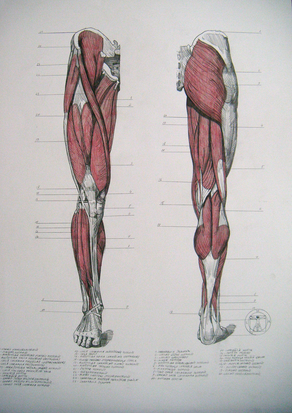
Muscles of legs. Front and back by reinisgailitis on ... from img00.deviantart.net It is responsible for extension,adduction, and (medial) internal rotation of the shoulder joint. Back view of muscles, skeleton, organs, nervous system. The muscle fibers' highly specialized structure enables the muscles to relax and contract to produce movement. 12 photos of the muscles labeled front and back. Aalso known as the six pack, is a paired muscle running vertically on each side of the front wall of the abdomen. Labeled viral infection explanation scheme. Vector illustration informative medical scheme. Given that muscles make movement happen, each muscle will create a certain movement around a joint.
Labeled anatomical ear structure scheme.
Labeled anatomical ear structure scheme. Triceps, biceps, pectoralis major, quadriceps , hamstrings, gluteus maximus , abdominals, deltoid, latissimus dorsi, external obliques, gastrocnemius , tibialis anterior. The muscles extend from the tubercles of the ribs behind, to the cartilages of the ribs in front, where they end in thin membranes, the external intercostal membranes. The superficial back muscles are the muscles found just under the skin. Your deltoid muscle at your shoulder has a front, middle, and rear part to it. Related posts of muscles labeled front and back. Want to learn more about it? Vector illustration informative medical scheme. There are two parallel muscles. The muscle fibers' highly specialized structure enables the muscles to relax and contract to produce movement. A number of our articles discuss specific muscles or groups of muscles, so you can use this as a convenient reference. The external intercostal muscles, or external intercostals (intercostales externi) are eleven in number on both sides. More specifically, this beautifully illustrated anatomy chart includes neck and shoulders, multiple views of the back and spine, and frontal views of each muscular extremity of the human body.
Virus disease symptoms and spreads infographic. There are two parallel muscles. The muscles of the anterior of the forearm are generally divided into two groups: Want to learn more about it? Click on the labels below to find out more about your muscles.
Anatomy- Muscle Actions - Biology 235 with Stern at San ... from classconnection.s3.amazonaws.com By doing these exercises, your shoulders will also improve in their overall. Click on the labels below to find out more about your muscles. Human muscle system, the muscles of the human body that work the skeletal system, that are under voluntary control, and that are concerned with movement, posture, and balance. Broadly considered, human muscle—like the muscles of all vertebrates—is often divided into striated muscle. This labeled human muscular system chart illustrates the major muscle groups in the back (posterior) view and the front (anterior) view. The external intercostal muscles, or external intercostals (intercostales externi) are eleven in number on both sides. A back muscle that pulls the arm down and back. Back view of muscles, skeleton, organs, nervous system.
The trapezius originates from the skull and spine of the.
The trapezius is the most superficial muscle of the back and forms a broad flat triangle. Each of your muscles is made up of thousands of thin, long, cylindrical cells called muscle fibers. Labeled viral infection explanation scheme. The anterior muscles of the torso (trunk) are those on the front of the body, including the muscles of the chest, abdomen, and pelvis. Leg muscle anatomical structure, labeled front, side and back view diagrams. Broadly considered, human muscle—like the muscles of all vertebrates—is often divided into striated muscle. The superficial back muscles are the muscles found just under the skin. Learn the muscles of the leg fast with these quizzes, diagrams and labeling exercises : Skeletal muscle groups front and back. There are two parallel muscles. Human muscle system, the muscles of the human body that work the skeletal system, that are under voluntary control, and that are concerned with movement, posture, and balance. The external intercostal muscles, or external intercostals (intercostales externi) are eleven in number on both sides. Attachments, nerve supply well there are lot of muscles on back and every muscle is trained differently.
The trapezius is the most superficial muscle of the back and forms a broad flat triangle. The muscles of the anterior of the forearm are generally divided into two groups: Labeled tick bite infection symptoms scheme. Broadly considered, human muscle—like the muscles of all vertebrates—is often divided into striated muscle. The muscle fibers' highly specialized structure enables the muscles to relax and contract to produce movement.
Muscles of legs. Front and back by reinisgailitis on ... from img00.deviantart.net Labeled anatomical ear structure scheme. The biggest muscle is lats muscle, then there are traps muscle. Learn the muscles of the leg fast with these quizzes, diagrams and labeling exercises : Rotator cuff muscle with anatomical posterior and anterior view expample. This labeled human muscular system chart illustrates the major muscle groups in the back (posterior) view and the front (anterior) view. Label muscles front and back view. Text and images from slide. C rnrceps brachn l unssimus dorsi k.
What do you prefer to learn with?
Front view of woman's thigh and knee muscles with names. What do you prefer to learn with? Human muscle system, the muscles of the human body that work the skeletal system, that are under voluntary control, and that are concerned with movement, posture, and balance. Each of your muscles is made up of thousands of thin, long, cylindrical cells called muscle fibers. The muscle fibers' highly specialized structure enables the muscles to relax and contract to produce movement. This labeled human muscular system chart illustrates the major muscle groups in the back (posterior) view and the front (anterior) view. By doing these exercises, your shoulders will also improve in their overall. Want to learn more about it? It is responsible for extension,adduction, and (medial) internal rotation of the shoulder joint. Label muscles front and back view. Text and images from slide. Male muscular system, full anatomical body diagram with muscle scheme, vector illustration educational poster. These muscles are able to move the upper limb as they originate at the vertebral column and insert onto.
Tags
julien nguyenart Diposting : Jumat, 12 Februari 2021, Februari 12, 2021